
Chiangmai Dentist and Chiangmai Dental specialists By Dental 4 You Clinic.
Leading dentist, specialist, cosmetic dentist, dental implant in Thailand.
Oral surgery
Tooth Extraction
There are a number of reasons why a tooth extraction is needed:
-
the tooth is severely decayed
-
advanced periodontal disease ("gum disease")
-
the tooth may be broken such that it cannot be repaired
-
other teeth may need removal because they are poorly positioned in the mouth (such as impacted wisdom teeth)
-
preparation for orthodontic treatment ("braces")
Procedure for Tooth Extraction
1. First Evaluation and diagnosis
-
An oral examination to determine if a tooth extraction is really warranted
-
X-ray(s) may be taken of the tooth to evaluate both the internal aspects of the tooth, the tooth root and bone
-
Relevant medical histories are recorded. Do ensure that you report to your dentist any problems with any previous tooth extractions, bleeding problems, medical conditions or allergies to medications and supplements
2. Site tooth preparation
-
Local anesthetize is given to "numb up" the tooth, jawbone and surrounding gums
3.Tooth removal
Recovery Expectations
For simple normal tooth extraction, the patient is simply sent home and the tooth site left to heal. Bleeding should stop after the first few hours. You may follow your regular daily activities, avoiding excessive exertion typed of activities such as exercising or sunbathing.
Postoperative care Instructions after Tooth Extractions
-
Bite the gauze firmly for a full 1-2 hour to stop bleeding. If bleeding persists, change to new gauze and continue biting firmly until the bleeding stops
-
Do not use mouthwash for 24 hours after oral surgery
-
If mild bleeding occurs, hold cold salt water in the mouth until it warms to body temperature
-
Do not rinse for 12 hours
-
After 12 hours you may rinse with a solution of tea spoonful of salt in a glass of warm water
-
Brush your teeth as usual, but do not brush the wound
-
Take only soft, non-spicy and cold foods, if possible, for 2- 3 days
-
Avoid smoking and alcohol
-
Mild pain can be controlled with pain relieve medications as directed by your dentist
Effects of Missing Teeth
When a tooth is missing its neighboring teeth will tend to shift, sometimes significantly, which in turn can have a major impact on your dental health. Even the removal of a single tooth can lead to problems related to your chewing ability, problems with your jaw joint, and the problem of a possibility that the surrounding teeth of predispose the teeth may shifted. Look for Benefits of Prosthetics at Prosthodontic Dentistry.
The Wisdom Tooth Removal
Wisdom teeth are third molars. Normally people have three permanent molars that develop in each quadrant of the mouth. The third molars usually will try to grow in at around age 18 to 20 years.
Wisdom teeth are actually no different than any other tooth except that they are the last teeth to erupt. They are just as useful as any other tooth if they grow in properly, have a proper bite relationship, and have healthy gum tissue around them. Unfortunately, this does not always happen.
When wisdom teeth are not erupting into the mouth properly, they are referred to as impacted teeth. A dentist must examine a patient's mouth and corresponding x-rays to determine if the teeth are impacted or will not erupt properly. Impacted teeth may cause problems. Here are several common problems that occur when the impacted wisdom teeth are not removed:
-
bacteria and plaque build-up
-
decay of adjacent teeth
-
formation of cysts (a fluid- filled sac) or tumors from follicle
-
tumor development
-
infection
-
jaw and gum disease
Erupted wisdom teeth may need to be removed. The dentist may recommend wisdom tooth removal if wisdom tooth:
-
interferes with the bite
-
non-functional
-
badly decayed
-
involved with or at risk for periodontal disease
-
interferes with restoration of an adjacent tooth
Every case is different and only your dentist can determine if there is a reason for you to have a tooth removed.
Symptoms of Wisdom Tooth Removal
The following symptoms may indicate that the wisdom teeth have erupted and surfaced. However, each individual may experience symptoms differently. Symptoms may include:
-
pain
-
infection in the mouth
-
facial swelling
-
swelling of the gum line in the back of the mouth
Many oral health specialists will recommend removal of the wisdom teeth, (when the roots are approximately formed, or three-fourths developed, usually in the adolescent years), as early removal will help to eliminate problems, such as an impacted tooth that destroys the second molar. Third molar impaction is the most prevalent medical developmental disorder.
Procedure for Wisdom Tooth Removal
1. First Evaluation and diagnosis
-
An oral examination to determine if a tooth extraction is really warranted
-
X-ray(s) may be taken of the tooth to evaluate both the internal aspects of the tooth, the tooth root and bone
-
Relevant medical histories are recorded. Do ensure that you report to your dentist any problems with any previous tooth extractions, bleeding problems, medical conditions or allergies to medications and supplements
2. Site tooth preparation
-
Local anesthetize is given to "numb up" the tooth, jawbone and surrounding gums
-
Wisdom tooth removal
-
Connective tissue between the tooth and the bone are removed
3. Wisdom Tooth is removed
-
Stitch up to close surgical site
Recovery Expectations
For simple normal tooth extraction, the patient is simply sent home and the tooth site left to heal. Bleeding should stop after the first few hours. You may follow your regular daily activities, avoiding excessive exertion typed of activities such as exercising or sunbathing.
Postoperative care Instructions after Tooth Wisdom Extractions
-
Use cold compress for 12-24 hours right after the surgery
-
Bite the gauze firmly for a full 1-2 hour to stop bleeding. If bleeding persists, change to new gauze and continue biting firmly until the bleeding stops
-
Do not use mouthwash for 24 hours after oral surgery
-
If mild bleeding occurs, hold cold salt water in the mouth until it warms to body temperature
-
Do not rinse for 12 hours
-
After 12 hours you may rinse with a solution of tea spoonful of salt in a glass of warm water
-
Brush your teeth as usual, but do not brush the wound
-
Take only soft, non-spicy and cold foods, if possible, for 2- 3 days
-
Avoid smoking and alcohol
-
Mild pain can be controlled with pain relieve medications as directed by your dentist
Bone Grafts
Bone grafting procedures are usually necessary if there is not enough bone available to place dental implants or if any vital anatomy is in the way.
Today, bone grafting procedures have become almost an integral part of implant reconstruction. In many instances, a potential implant site in the upper or lower jaw does not offer enough bone volume or quantity to accommodate a Root form Dental Implant of proper size or in the proper place. This is usually a result of bone resorption that has taken place since one or more teeth (if not all) were lost. Bone Grafting procedures usually try to re-establish bone dimension, which was lost due to resorption.
Many years ago the lack of bone posed a considerable problem and sometimes implant placement was impossible because of that. Today, however, we have the ability to grow bone where needed. This not only gives us the opportunity to place implants of proper length and width (and for Root form Implants we always try to go for as long and wide as possible), it also gives us a chance to restore the esthetic appearance and functionality better.
Our clinic use bone grafting and tissue regeneration products from Osteohealth Company .The products of this company have been used by dental professionals throughout the world. The company's overriding goal is to develop and distribute, with professionalism and integrity, products of the highest quality that have been scientifically and clinically proven to enhance patient care.
Bio-Oss® Natural Bone Grafting Material
Bio-Oss® is a natural, osteoconductive bone substitute that promotes bone growth in periodontal and maxillofacial osseous defects. It consists of the mineral portion of bovine bone. Bio-Oss® provides the body with a matrix for bone cell migration and is integrated into the natural physiologic remodeling process.
Bio-Oss® natural bone mineral offers predictable results which have been proven through years of clinical experience and extensively documented in published scientific literature.
Bio-Oss®: Morphology like human bone
Bio-Oss® was developed as an ideal bone substitute that would replicate the structure of autogenous bone. Each step in the development of Bio-Oss® was conducted with this goal in mind, resulting in a matrix that is very similar in physical and chemical composition to human bone.
The trabecular architecture and fine crystalline structure of the natural bone in Bio-Oss® are preserved through a patented manufacturing process, resulting in an exceptional osteoconductive matrix.
High similarity of Bio-Oss® to human bone
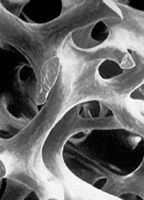
Bio-Oss® Cancellous Structure

Bio-Oss® - small and compact nanocrystals
similar to human bone. (TEM 100,000x)

Autogenous Bone Structure

Human bone - small and compact
natural apatite crystals (TEM 100,000x)
Process of natural bone regeneration with Bio-Oss®
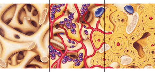
-
Clot stabilization facilitated by Bio-Oss® interconnecting macro and micropores.
-
Revascularization, migration of osteoblasts (purple) and in-growth of woven bone (yellow) is enhanced by Bio-Oss® scaffolding.
-
Lamellar bone and Bio-Oss® are successfully integrated after approximately six months. Bio-Oss® is included in the natural physiologic remodeling process (osteoclasts - Blue).
Advantages of Bio-Oss®
-
Superior handling characteristics made possible by the large hydrophilic inner surface area similar to human bone.
-
Promotes revascularization and clot stabilization, due to its interconnecting macro and micropores.
-
Facilitates bone formation by providing an exceptional osteoconductive scaffolding which results from the retention of the natural porous architecture and trabeculation of human cancellous bone.
-
Effective space maintenance, and when integrated, provides mechanical strength and stiffness due to retention of the natural mineral content.
-
Optimal integration with patients own bone aided by a chemical composition analogous to human bone with fewer hydroxyl and more carbonate groups than most synthetic materials.
-
Bio-Oss® is integrated during the natural remodelling process of the human bone and slowly resorbed due to small crystallite size which is comparable to human bone.
-
Effective bone regeneration that has been clinically and scientifically proven for more than 15 years.
-
Bio-Oss® prevents newly formed bone from rapid resorption and leads to a long-term preservation of the bone volume.
Bio-Gide® Resorbable Bilayer Membrane
Bio-Gide® consists of highly purified collagen types I and III (porcine origin). The membrane is highly biocompatible and supports wound healing. The three-dimensional, natural fiber structure promotes cell adhesion, serves as a matrix for soft tissue support and provides a barrier to the ingrowth of overlying soft tissue into underlying bony defects.
Experience shows there is low risk of postoperative membrane exposure. Should wound dehiscence occur, soft tissue healing and closure of the dehiscence defect usually follows.
The barrier function of Bio-Gide® lasts for several months prohibiting soft tissue ingrowth into the bony defect facilitating undisturbed bone regeneration. Resorption takes place enzymatically and without irritation.
The native collagen fibers of Bio-Gide® are strongly hydrophilic. As a result, once moistened, the membrane adapts very well to the surgical site.
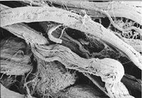
Bio-Gide® bilayer design, cross section (SEM 100x)

High similarity of Bio-Gide® to human collagen membrane
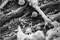
Bio-Gide® - natural collagen structure (SEM 2000x)
The Bio-Gide® membrane consists of collagen types I and III which have been prepared to result in a bilayer membrane. The dense superior membrane layer, which faces the soft tissue, is cell occlusive and prevents invasion of soft connective tissue cells into the membrane protected space. The porous inferior layer, which faces the bony defect, consists of loosely arranged collagen fibers which act to stabilize the clot and enable bone cells to become integrated into the membrane.
The dense cell-occlusive layer is a barrier to soft tissue ingrowth and serves as a soft tissue scaffold (blue arrows). Granulation cells adhere to the natural collagen surface.
The porous side of the membrane is an open-pore, three-dimensional collagen matrix, which promotes cell integration (white arrows).
Human Schneiderian membrane dissolved on the bony side.
SEM view 2000x (Prof. K. Brenner, Anatomy Institute, LMU Munich)
Advantages of Bio-Gide®
Material
Properties
Results
Tear-proof fibrous tissue structure
May be trimmed to desired size/shape and subsequently fixed in place either by adherence or fixation with pins/sutures
Easy to handle and perfect adaptation to the defect
Matrix with a bilayer structure
The smooth cell-occlusive layer forms a natural barrier against soft connective tissue cells
The porous layer favors bone tissue integration
Long term undisturbed bone regeneration
Matrix with a bilayer structure
Accelerate bone cell adherence and blood clot stabilization
Resorption without irritation of the tissue by acid formation
Favorable soft tissue
healing Single step surgery
Maxillary sinus lift
A sinus lift (a sinus augmentation) is surgery that adds bone to your upper jaw in the area of your molars and premolars to make it taller. The bone is added between your jaw and the maxillary sinuses, which are on either side of your nose. To make room for the bone, the sinus membrane has to be moved upward, or "lifted." A sinus lift usually is done by an oral and maxillofacial surgeon or a periodontist. It can happen that there is no longer enough bone in the molar area of the maxilla in the direction of the floor of the sinus because of the bone atrophy after tooth loss. As bone augmentation in this region is possible only with difficulty, a method was developed in which the floor of the sinus is raised and bone is inserted into the cavity produced, without injuring the mucosa of the sinus, which leads to an effective increase in the bone in the molar maxillary region. This operation is called a sinus lift. This operation can be carried out under local anaesthesia and is done through the mouth so that there are no scars on the face. The sinus lift operation can often be done at the same time as the insertion of dental implants. The bone material which is inserted corresponds to that described already for bone augmentation.
A sinus lift is done when there is not enough bone in the upper jaw, or the sinuses are too close to the jaw, for dental implants to be placed. There are several reasons for this:
-
Many people who have lost teeth in their upper jaw — particularly the molars teeth — do not have enough bone for implants to be placed. Because of the anatomy of the skull, the back of the upper jaw has less bone than the lower jaw.
-
Once teeth are gone, bone begins to be resorbed (absorbed back into the body). If teeth have been missing for a long time, there often is not enough bone left to place implants.
-
The maxillary sinus may be too close to the upper jaw for implants to be placed. The shape and the size of this sinus varies among individuals. In addition, the sinus can get larger as you age.
-
Bone may have been lost because of periodontal (gum) disease.
Dental implants have gained popularity for treating edentulism, but some patients develop jaw atrophy, which leaves insufficient bone for implants. To treat these patients, the sinus lift procedure, which augments bone, was developed. Altered anatomy from this procedure has an unusual radiographic appearance, confusing those unfamiliar with it. We describe the sinus lift procedure and its radiographic appearance.

Procedure
The oral surgeon will cut the gum tissue near your premolars and molars. The tissue is raised, exposing the bone. A small, oval window is opened in the bone. The membrane lining the sinus on the other side of the window separates your sinus from your jaw. This membrane is gently pushed up and away from your jaw. Granules of bone-graft material are then packed into the space where the sinus was. The amount of bone used will vary, but usually several millimeters of bone is added above the jaw.
Once the bone is in place, the tissue is stitched closed. Your implants will be placed four to nine months later, depending on the graft material that was used. This allows time for the grafted material to mesh with your bone.